
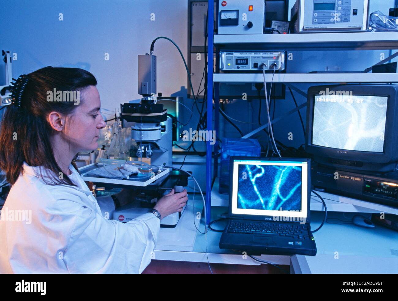
Microcirculation research on a mouse using the cell viZio imaging system. The cell viZio, which was developed by Mauna Kea Technologies, France, allow
RMID:Image ID:2ADG96T
Image details
Contributor:
Science Photo LibraryImage ID:
2ADG96TFile size:
50.3 MB (1.7 MB Compressed download)Releases:
Model - no | Property - noDo I need a release?Dimensions:
5050 x 3479 px | 42.8 x 29.5 cm | 16.8 x 11.6 inches | 300dpiDate taken:
14 October 2004Photographer:
PHILIPPE PSAILA/SCIENCE PHOTO LIBRARYMore information:
Microcirculation research on a mouse using the cell viZio imaging system. The cell viZio, which was developed by Mauna Kea Technologies, France, allows microscopic images of tissue to be taken in living animals. It allows the flow of blood cells (haemodynamics) and structures of blood vessels to be studied with minimum invasion. The system consists of a long tube containing several thousand flexible optical fibres. At the tip of the tube is a very thin probe (350 microns diameter), which can enter the tissue. A laser shines on the tissue and relays the digital microscopic image to a computer, which displays the image on a monitor (lower right).

